3-D Bioprinted Tissue Model Gives Hope for Improving Drug Absorption

Study in a Sentence: An international group of scientists collaborated to develop a vascularized 3-D bioprinted liver tissue that includes an endothelial cell-layered channel network, allowing researchers to better observe the drug administration process.
Healthy for Humans: Adding the endothelial layer to the vascularized 3-D human tissue provided researchers with a model that more accurately mimics the structure and function of a human liver. Due to this new complexity, researchers found the endothelial layer delayed the drug diffusion response, because it took time for a compound to permeate the endothelial layer, making the model a more accurate representation of patients’ livers.
Redefining Research: Researchers will use the bioprinted tissue to study drug diffusion in an effort to improve drug absorption. The model also allows researchers to use a patient’s own cells, providing a more advanced and personalized platform for testing whether a drug is likely to be safe or toxic for a particular patient. Often, differences between patients in the effectiveness or toxicity of a drug are due to differences in the rate of drug absorption.
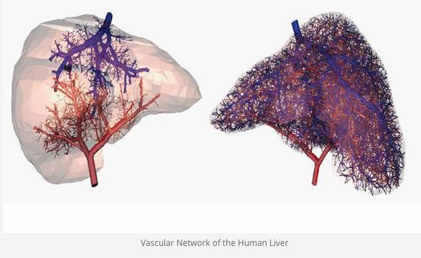
This image shows a vascular network of the human liver. Photo credit: 3DPrint.Com
References
- Massa S, Sakr MA, Seo J, et al. Bioprinted 3D vascularized tissue model for drug toxicity analysis. Biomicrofluidics. Published online August 2017. doi: http://dx.doi.org/10.1063/1.4994708.


